Tampa General Hospital's in-house 3D Medical Visualization and Printing Lab translates detailed clinical images—such as computed tomography (CT) and magnetic resonance imaging (MRI) scans—into patient-specific, three-dimensional devices, such as anatomical models and guides that help healthcare teams see complex anatomy in a way that a flat scan simply cannot show.
Our 3D-printed models support both surgical planning and communication, allowing surgeons to closely evaluate anatomical features and anticipate potential challenges long before entering the operating room. Patients and their families can also see and touch a physical representation of the anatomy involved in their care—making it easier to ask questions and understand the treatment plan.
About 3D Medical Visualization and Printing
Using standard medical imaging and cutting-edge software, the 3D medical visualization and printing process converts digital anatomy into highly accurate physical models. This approach, known as additive manufacturing, builds lifelike objects layer by layer, allowing the model to capture therapeutically relevant details, such as tumors, injuries and congenital differences.
Depending on the clinical need, models can be produced in medical-grade materials ranging from rigid, bone-like plastics to flexible, tissue-mimicking polymers. This same technology can also be used to create procedure-support tools, including biocompatible, sterilizable cutting and positioning guides designed to conform to a patient's unique body structures.
When a surgeon is planning a technically demanding procedure, explaining it to the patient, or rehearsing surgical techniques, an exact replica of the patient's anatomy can be an invaluable tool. TGH and USF Health are proud to be the first in the Tampa Bay area—and among the first in the nation—to bring 3D medical visualization and printing into clinical practice.
Why Choose Tampa General's 3D Lab
Nationally-Recognized for Excellence
TGH and USF Health are nationally recognized for excellence in patient safety, clinical outcomes, and medical leadership. As a prominent academic health system, we pair specialized expertise with leading-edge technology to deliver high-quality, evidence-based care. Our focus on continuous improvement strengthens our performance across specialties while ensuring our patients can benefit from new approaches and resources designed to support better health and quality of life.
Central to our reputation as a regional leader in point-of-care additive manufacturing is our integration of 3D medical visualization and printing directly into the hospital setting. By providing these services on-site, we enable close collaboration among clinicians, researchers and technical specialists, powering the kind of close collaboration that produces better outcomes. It is a deliberate choice—and one that reflects how seriously we take the promise of bringing this technology to bear at the point of care.
Four Pillars of Our Work
Everything we do in the lab connects back to four main pillars where 3D medical visualization and printing drives real, measurable change. Each pillar reflects both our commitment to innovation and our approach to solving problems that matter to patients, surgeons, educators, and researchers.
Clinical
Patient-specific anatomical models and surgical guides go directly from imaging to the operating room—on-site, with no third-party delays. Our team works hand-in-hand with surgeons and clinicians on cases ranging from presurgical planning to complex reconstructions, translating images into physical tools that support better preparation and more confident execution.
Our lab provides individualized collaboration for a wide range of specialties, including radiation oncology, cardiology, vascular surgery, pulmonology, orthopedic surgery, ENT, OB-GYN, plastic surgery, neurosurgery and transplant surgery, among others.
Research
Our lab is an active partner in clinical research and medical device development. From rapid prototyping to technology evaluation, we work with USF Health and other collaborators to push the boundaries of what point-of-care additive manufacturing can do—turning clinical questions into testable, tangible solutions.
We are also working on the visualization side, developing software to improve better understanding from the final user, adding value to the process.
Education
Through our partnership with USF Health, the lab produces anatomical models for simulation, procedural rehearsal, and resident training. Clinicians and students gain hands-on experience with real anatomical complexity, including rare presentations and unusual geometries that no textbook illustration can fully capture.
Facilities
TGH's lab runs industrial-grade PolyJet, SLA/DLP, and FDM printing systems inside the hospital—not in a separate facility. That proximity matters. With that, we can help different areas create any replacement part the hospital needs.
World-Class Anatomic Modeling by Experts
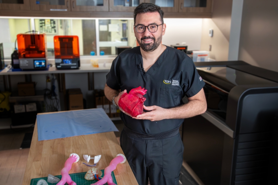
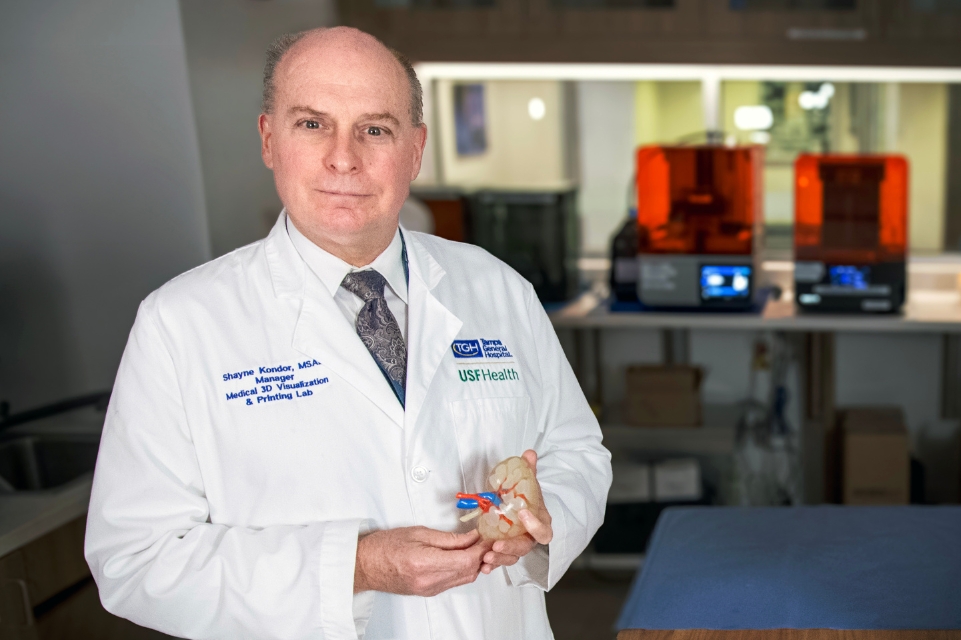
TGH's 3D medical visualization and printing lab brings together a multidisciplinary team comprised of biomedical engineers, specialized radiologists and surgeons. Key members include:
Our Comprehensive Point-of-Care Facility
For applications ranging from presurgical planning for severe traumatic injuries to highly detailed educational models for rare congenital conditions, TGH enables precise, patient-centered care through 3D medical visualization and printing. By transforming medical images into accurate three-dimensional visualizations—and, when needed, procedure-specific cutting and positioning guides—we help healthcare teams better understand each patient's distinct anatomy and prepare for the nuances of complex cases.
TGH's on-site lab brings engineering and clinical expertise together within the hospital environment, supporting a true bench-to-bedside approach where technical development and clinical application happen in close coordination. This integrated workflow helps simplify the path from imaging to procedural planning, enabling faster iteration and more efficient decision-making.
For patients, it supports greater procedural confidence and more precise surgical execution—reducing time in the operating room, improving treatment accuracy, and easing recovery. It is another expression of TGH's long-held focus on personalized care and clear communication with every patient we serve.
3D Lab Services & Capabilities
Working closely with clinicians, TGH's 3D lab team provides a range of services to transform medical imaging into practical tools that can be used to plan and deliver treatment or provide education and training. Our capabilities include:
- Virtual surgical planning (VSP) – Patient imaging can be used to create detailed digital models, allowing surgeons to evaluate anatomy, simulate procedural steps and refine the surgical approach before the first incision is made.
- High-fidelity anatomical models – Accurate physical replicas of patient anatomy can represent rigid structures, such as bone, as well as flexible soft tissues, supporting improved visualization, planning and communication.
- Custom biocompatible and sterilizable guides – Designed to match patient anatomy, procedure-specific guides can be manufactured using materials that can be taken directly into the operating room, helping to translate the surgical plan into precise execution.
- Medical device prototyping – Rapid development and evaluation of customized medical devices and components can promote clinical innovation, workflow optimization and iterative design.
- Education and simulation training models – Anatomical models can be created for hands-on training, procedural rehearsal and education for clinicians, medical students and interdisciplinary teams.
- Research and development – Collaborative projects can explore new applications of 3D visualization and printing to support clinical research, technology evaluation and the advancement of patient-focused solutions.
State-of-the-Art Additive Manufacturing
TGH is committed to providing our patients with access to advanced technologies that support individualized treatment and informed decision-making. Toward that end, we have equipped our 3D Medical Visualization and Printing Lab with several industrial-grade additive manufacturing systems, including:
- Photopolymer jetting (PolyJet) – PolyJet uses a high-resolution process that jets and cures liquid photopolymer resin in ultra-thin layers. Particularly well-suited for creating intricate anatomical models, this system can combine different material properties (such as rigid and flexible) within the same print.
- Stereolithography/digital light processing (SLA/DLP) – SLA and DLP are resin-based printing methods that use light to cure liquid photopolymer into solid layers. SLA typically uses a laser, while DLP uses a projected light image. These technologies are valued for producing smooth surface finishes and fine detail, making them useful for creating precise anatomical models and customized procedural tools.
- Fused deposition modeling (FDM) – The FDM process creates distinct parts by heating and extruding a thermoplastic filament through a nozzle, building the model layer by layer. Often used for durable models and prototypes, this technology may be an efficient option for larger prints or components when strength and practicality are priorities.
When developing 3D anatomical models, different clinical applications may call for different levels of rigidity, flexibility, transparency and tactile "feel." To meet the specific goals of each case, our team selects from a broad range of materials designed to replicate key structural properties and highlight the details that matter most for planning and communication. These include:
- Rigid, bone-like materials – Can be used to represent dense structures, such as bone for orthopedic and craniofacial applications, where accurate geometry and stable handling are important for evaluating alignment, defects or reconstruction needs.
- Flexible, tissue-mimicking photopolymers – Can be used to model soft tissue anatomy and multifaceted relationships between structures—such as how a lesion, defect or another abnormality interacts with the surrounding tissues—while providing a realistic sense of pliability and spatial context.
- High-stability & resilient materials (Tough 1500) – This is our "workhorse" material for the behind-the-scenes parts of a project. Because it provides high mechanical stability, it is incredibly strong and holds its shape under pressure without being brittle. We use this for non-clinical applications, such as creating custom supports, specialized racks, or sturdy fixtures that hold other parts in place during the manufacturing process. It's the perfect choice when we need a part that can be handled daily and stay "tough" without breaking.
- Sterilizable & biocompatible materials (BioMed Clear) – This material is specifically designed for medical safety. It is biocompatible, meaning it has been officially tested for safe contact with the human body, and it is durable enough to be sterilized for use in professional clinical environments.
These state-of-the-art platforms and materials allow our team to produce highly detailed, patient-matched models and procedural-support tools with heightened accuracy and consistency.