Innovation - Advanced 3D Imaging
Published: Feb 17, 2023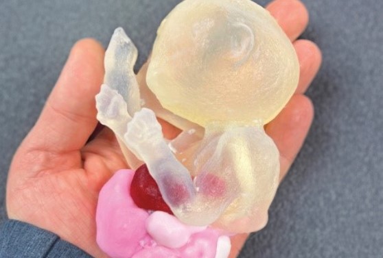
Advanced 3D Printing Transforms Complex, Specialized Procedures at TGH
In 2011, Tampa General Hospital and the USF Health Morsani College of Medicine Department of Radiology established one of the first clinical 3D laboratories in Florida, specifically to print replicas of human hearts to aid in complex surgical procedures. As an international test site for the technology, TGH has continued to expand those capabilities to include 3D printing applications in cardiology, orthopedics, gynecology, pediatric surgery and maternal-fetal medicine.
The TGH and USF Health Radiology’s Division of 3D Clinical Applications is one of the most advanced point-of-care printing labs in the nation, creating 3D models that are accurate in size and shape to within one one-hundredth of an inch. The models are biomimetic and bio-compatible — giving physicians near-perfect replicas to aid in surgical planning and procedural practice.
Working in collaboration with the Radiology 3D team, gynecologists at TGH and USF Health are using 3D modeling to improve care for patients with deep endometriosis, taking the guesswork out of which approach to take in providing treatment for patients suffering immense pain and discomfort.
While many hospitals may outsource this type of printing service, surgeons at TGH and USF Health have access to 3D models and prints more quickly, giving them more time to determine the best approach and advancing the quality of care.
3D Printing Improves Fetal and Pediatric Medicine at TGH
Surgeons use this technology for high-risk procedures to treat babies in utero. For example, EXIT, short for Ex Utero Intrapartum Treatment, is a highly complex procedures used to treat babies in utero with life-threatening conditions that must be addressed before delivery.
TGH is one of the few hospitals in Florida with the expertise to perform this complex procedure and has been performing the EXIT procedure since 2009. With the introduction of 3D printing, surgeons at TGH are better able to plan and execute the operation with more confidence, and less time in the OR, which significantly improves outcomes for the mother and baby.
Biomimetic models of fetuses in utero can also be used to show parents the nature of their babies’ severe deformities in a way that cannot be seen in ultrasound. Not only does it aid in decision-making, but it also allows surgeons to reassure parents that they’ve come to the right place. For pediatric surgeons treating cranial facial deformities, 3D printing of the baby’s skull improves procedural planning and practice. As a result, surgeons spend less time performing surgeries, patients require less anesthesia, and the subsequent risks decrease.
Home to First Dedicated 3D Radiology Residency Training in the U.S.
3D modeling at TGH draws some of the most difficult-to- treat cases in the region and has helped establish the first 3D training within a radiology residency in the nation. Over 30 residents participate in a month-long 3D radiology training rotation at TGH, which increases the number of specialists in the field. TGH and USF Health are committed to making a meaningful contribution to next-generation radiology and 3D modeling nationwide.